Computed Tomography (CT) Scan
Jump to navigation
Jump to search
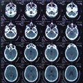
A Computed Tomography (CT) Scan is a medical image based on X-ray scans taken from multiple angles.
- Context:
- It can (typically) create detailed cross-sectional images of the body.
- It can (typically) create detailed three-dimensional cross-sectional images.
- It can (typically) be created by a CT Scanner ...
- It can (typically) involve Tomographic Reconstruction Algorithms.
- It can (often) enable differentiation between tissues in the body with different Radiodensity Levels (which is essential for identifying abnormalities such as tumors, blood clots, and fractures).
- It can range from being a Standard CT Scan, which uses a moderate amount of X-Ray Radiation, to a Low-Dose CT Scan, which reduces radiation exposure but may offer less detail in images.
- It can utilize contrast materials like Iodine-Based Contrast Agents to enhance image clarity, particularly for blood vessels and internal organs.
- It can be contraindicated in certain scenarios due to its use of Ionizing Radiation, which poses potential risks such as cancer from excessive exposure.
- ...
- Example(s):

- a CT Scan of the Brain that can detect strokes, tumors, or other neurological issues by providing detailed images of brain tissue.
- a CT Scan of the Abdomen that helps diagnose problems in the liver, kidneys, and other abdominal organs.
- ...
- Counter-Example(s):
- Magnetic Resonance Imaging (MRI)s, which use magnetic fields and radio waves instead of X-rays and are preferred in scenarios where radiation exposure must be minimized.
- Ultrasound Imaging, which uses sound waves and is often used for real-time imaging processes such as pregnancy scans.
- ...
- See: Kidney Problems, X-Ray, Tomography, Geometry Processing, Three-Dimensional Space, Radiography, Axis of Rotation, Medical Imaging, Diagnosis, Industrial Computed Tomography Scanning, Positron Emission Tomography, Single-Photon Emission Computed Tomography.
References
2022
- (Wikipedia, 2022) ⇒ https://en.wikipedia.org/wiki/CT_scan Retrieved:2022-5-10.
- A CT scan or computed tomography scan (formerly known as computed axial tomography or CAT scan) is a medical imaging technique used in radiology (x-ray) to obtain detailed internal images of the body noninvasively for diagnostic purposes. The personnel that perform CT scans are called radiographers or radiology technologists. CT scanners use a rotating X-ray tube and a row of detectors placed in the gantry to measure X-ray attenuations by different tissues inside the body. The multiple X-ray measurements taken from different angles are then processed on a computer using reconstruction algorithms to produce tomographic (cross-sectional) images (virtual "slices") of a body. The use of ionizing radiation sometimes restricts its use owing to its adverse effects. However, CT can be used in patients with metallic implants or pacemakers, for whom MRI is contraindicated. Since its development in the 1970s, CT has proven to be a versatile imaging technique. While CT is most prominently used in diagnostic medicine, it also may be used to form images of non-living objects. The 1979 Nobel Prize in Physiology or Medicine was awarded jointly to South African-American physicist Allan M. Cormack and British electrical engineer Godfrey N. Hounsfield "for the development of computer-assisted tomography".